Arshavsky Laboratory
We are interested in molecular mechanisms underlying signal transduction, subcellular compartmentalization, and healthy state in vertebrate photoreceptor cells. Rod and cone photoreceptors are sensory neurons responsible for the detection and primary processing of information entering the eye in the form of photons of light. Photoreceptors capture photons, generate a second messenger signal, translate this signal into a change in electrical activity, and transmit this information to the secondary neurons in the retina through modulation of their synaptic release.
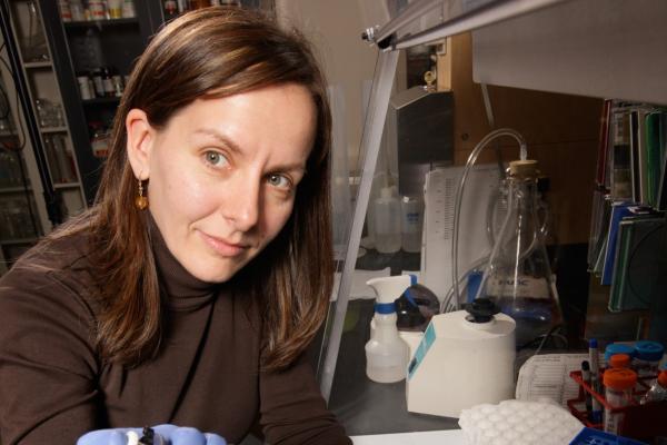
Bowes Rickman Laboratory
We are interested in the molecular mechanisms underlying the development of age-related macular degeneration (AMD). Currently our studies are focused on development and studies of animal models of AMD, AMD pathogenesis and pre-clinical studies of novel therapies for AMD.
Duke Reading Center
The Duke Reading Center is a comprehensive image reading center that specializes in reliable, timely, reproducible, and systematic analysis of images captured by many different retinal imaging technologies, including optical coherence tomography, fundus autofluorescence, color fundus photography, and fluorescein angiography. The Duke Reading Center is dedicated to quality, efficiency, and innovation by constantly improving and refining research protocols and embracing cutting-edge technology and research techniques.

Farsiu Laboratory
At the Vision and Image Processing (VIP) Laboratory at Duke University, our long-term goal is to improve the overall health and vision outcomes of at-risk patients with ocular and neurological diseases through earlier and better-directed therapy. To achieve this goal, we take advantage of recent advances in artificial intelligence (AI) and optics as an integrated technology to capture the highest quality ocular images.
Ferreira Laboratory
The focus of the Ferreira laboratory is to understand the integration of signaling and trafficking pathways and how of such pathways relay environmental cues across subcellular compartments and cellular systems in mouse and disease models. Central to our interests is the discovery of the role of such pathway networks in the modulation of aging and disease processes leading to neurodegeneration and other human maladies.
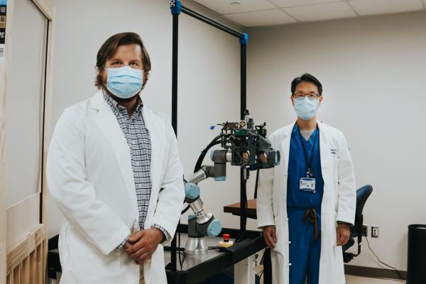
Kuo Laboratory
The Kuo laboratory is focused on developing the technologies necessary create digital images of the eye for clinical use. Our current projects include developing a “Whole Eye” scanner based on optical coherence tomography (OCT) and developing software to ensure the spatial accuracy of the scans. These scans will be used clinically to improve measurements of the eye in preparation for surgery and also to study how the back of the eye changes with myopia (near-sightedness).
Liton Laboratory
The long-term goal of our research program is elucidating the molecular mechanisms underlying the normal physiology and the pathophysiology of the outflow pathway. The outflow pathway is a complex tissue located in the anterior segment of the eye responsible of maintaining proper levels of intraocular pressure. Failure of this tissue with aging is associated with increase risk in developing Primary Open Angle Glaucoma, a blinding disease, affecting more than 70 million people worldwide. At present it is not known why this tissue fails with aging and in disease. To address this fundamental question, my laboratory has developed two major independent, but yet interconnected, research programs.
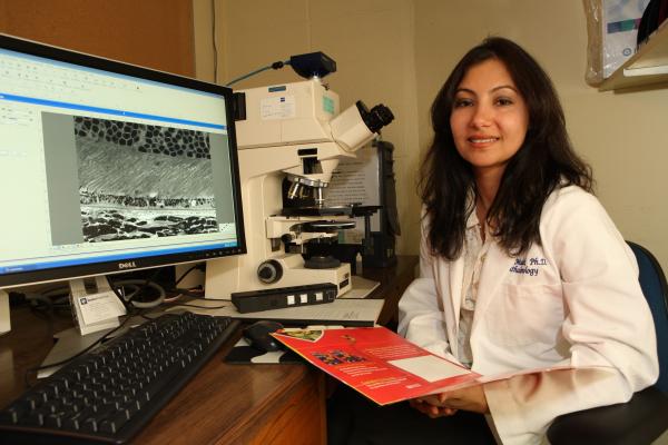
Malek Laboratory
Age-related macular degeneration (AMD) is the leading cause of central visual impairment amongst the elderly in the Western World and is becoming increasingly prevalent World-wide. Our lab is focused on understanding the cellular and molecular pathogenic mechanisms underlying the dry and wet forms of AMD. Our ultimate goals are to identify signaling pathways, which become dysfunctional in AMD; develop animal models, which present with features of the human disease; and identify potential therapeutic targets.
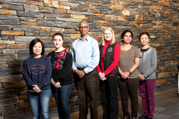
Rao Laboratory
Research in our laboratory is focused broadly on understanding involvement of the actin cytoskeleton and cell adhesive mechanisms in normal function and physiology, and the pathological processes that impact two ocular tissues: Lens and Trabecular meshwork.

Saban Laboratory
My broad research interests are the cellular and molecular mechanisms that contribute to pathogenic immunity in ophthalmic disease and vision loss. My studies are currently focused on dendritic cells (DC), a unique leukocyte population of antigen presenting cells required for both initiating and determining the type of immune response generated. These cells contribute to the maintenance of health versus immunity in ocular disease.
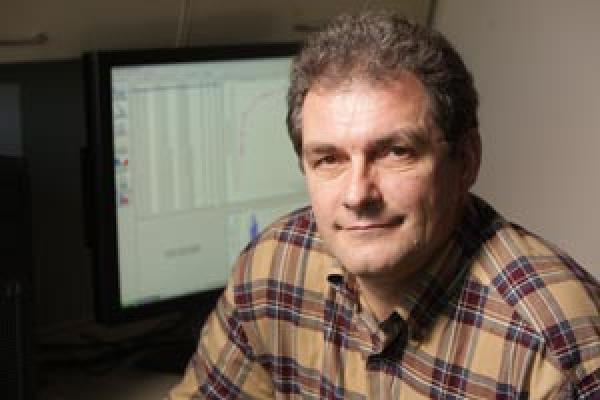
Skiba Laboratory
My research focuses on applying mass spectrometry based proteomics to study proteins in eye tissues, cells and sub-cellular compartments to understand mechanisms of vision. An important aspect of my research is to identify proteins in different compartments of retinal photoreceptor cells, their amount and modification status at different cell states defined by the light conditions, genotype, disease etc. This information can be valuable in understanding molecular mechanisms of vision and biology of the photoreceptor cell.
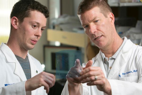
Stamer Laboratory
My laboratory studies the disease of glaucoma, the second leading cause of blindness in the United States, affecting nearly 3 million people (70 million Worldwide). The primary risk factor for developing glaucoma is ocular hypertension (high intraocular pressure, IOP). IOP is a function of aqueous humor moving into and out of the eye. Elevated IOP in glaucoma is a result of disease in the primary efflux route, the conventional outflow pathway, affecting proper drainage of aqueous humor.
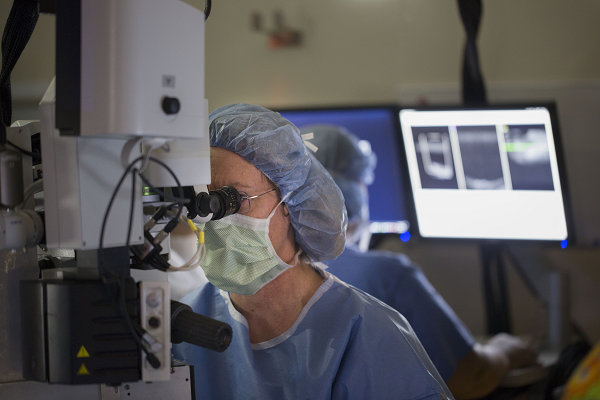
Toth - DARSI Lab
Duke Advanced Research in Spectral Domain OCT Imaging
Cynthia Toth, MD is an authority in the development and research/clinical application of ophthalmic optical coherence tomography (OCT) with more than 25-years of translational research and over 80 publications in this field out of her 200 peer-reviewed publications. She has been an effective translational researcher in this field.