
Unilateral Serous Macular Detachment Accompanying Cystoid Macular Edema After COVID-19 Vaccination
Authors
Navid Elmi Sadr MD*, Ramyar Hariri MD
Introduction
COVID-19 infection, caused by the SARS-CoV-2 virus, was the primary global concern for at least three years. The burden of the associated pandemic made the global health system face urgent calls for the development of a vaccine. In the relatively short period that COVID-19 vaccines have been in use, several ocular adverse events have been reported, including ophthalmic vein thrombosis, acute macular neuroretinopathy, multiple evanescent white dot syndrome, paracentral acute middle maculopathy, multifocal choroiditis, Vogt-Koyanagi-Harada disease, new-onset Graves' disease, central serous chorioretinopathy, uveitis, corneal graft rejection, scleritis, episcleritis, facial nerve palsy, and abducens nerve palsy, among others.1,2 Herein, we report an unusual case of unilateral serous macular detachment accompanied by cystoid macular edema (CME), which occurred in close temporal association with the administration of a second dose of the Sinopharm COVID-19 vaccine.
Case Report
A 67-year-old male presented with progressive, painless vision loss in his left eye, noticed approximately 48 hours before presentation. He had received the second dose of the Sinopharm COVID-19 vaccine 7 days before presentation to the clinic. He had no systemic symptoms except mild generalized myalgia for the first 2 days post-vaccination. His medical history included benign prostatic hyperplasia, hypertension, and ischemic heart disease. There was no previous history of ocular or systemic autoimmune or infectious diseases. He was taking warfarin, acetylsalicylic acid, losartan, hydrochlorothiazide, carvedilol, atorvastatin, tamsulosin, and finasteride.
His blood pressure at the time of examination was 135/85 mmHg. Best-corrected visual acuity was 20/20 in the right eye and counting fingers in the left eye. Slit-lamp biomicroscopy revealed mild nuclear cataracts in both eyes. Fundus examination revealed sensory retinal detachment in the macula of the left eye. The optic nerve head and retinal periphery appeared to be normal bilaterally.
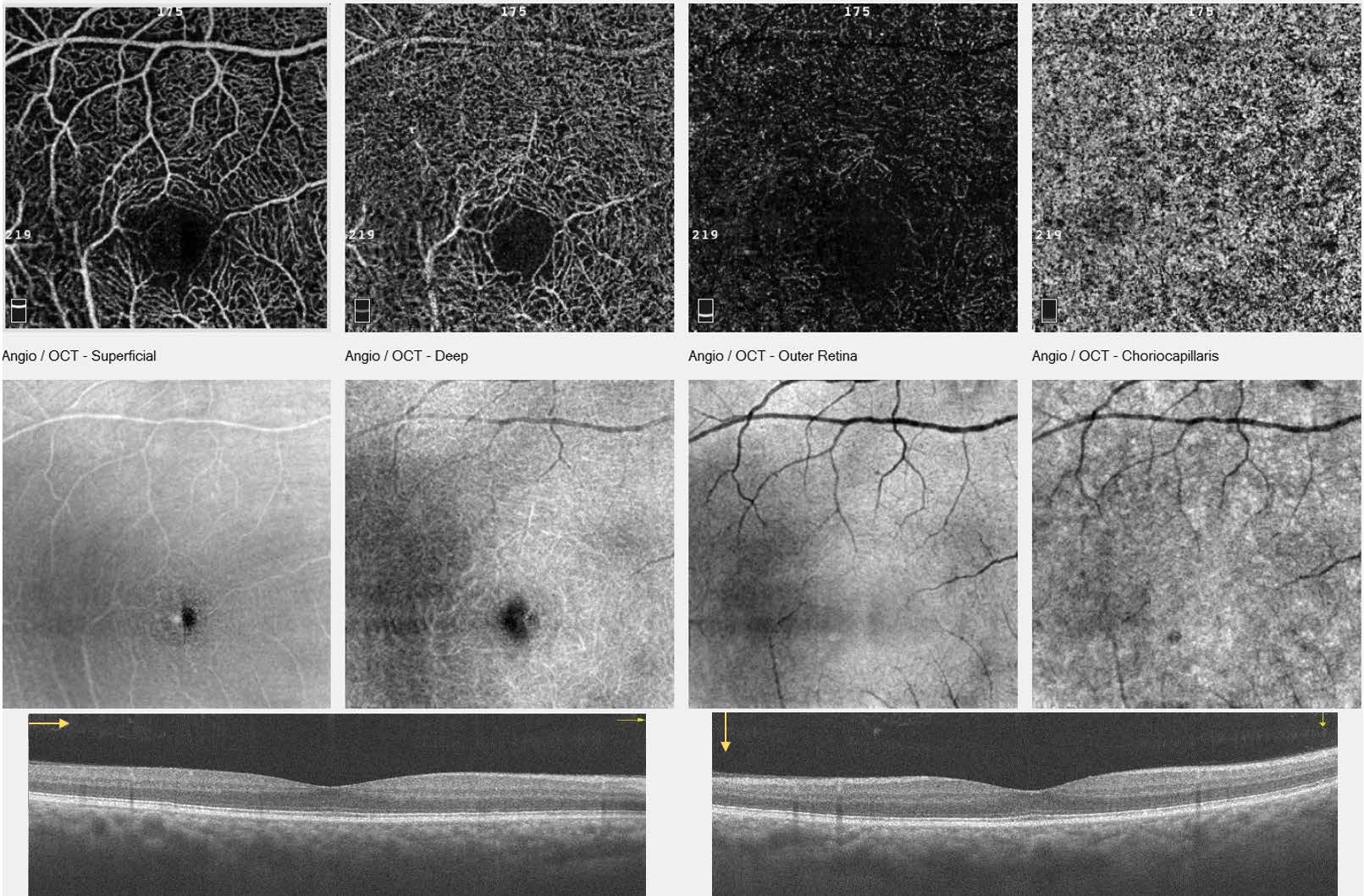
Spectral-domain optical coherence tomography angiography (SD-OCTA; Optovue, Fremont, CA) showed a large serous retinal detachment and CME with a lack of flow pixels in the choriocapillaris in the left eye (Figure 1). OCT of the right eye revealed normal foveal structures (Figure 2). Other fundus imaging modalities, including fundus photography, fluorescein angiography, and indocyanine green angiography, were not available at that time.

subretinal fluid (yellow asterisk) and cystoid hyporeflective spaces (red asterisk) in the macula. The en face
OCT angiography images (top row) showed no flow abnormalities in the superficial, deep, and outer retina.
Note that the lack of flow pixels in the choriocapillaris resulted from segmentation error caused by the presence
of intraretinal and subretinal fluid.
normal foveal structures.
A reverse transcription polymerase chain reaction test for COVID-19 using nasopharyngeal swab sampling was negative. Laboratory test results, including complete blood count, erythrocyte sedimentation rate, and Creactive protein levels, were within normal limits. Examination on days 3 and 7 revealed that the amount of subretinal fluid was gradually decreasing. Finally, on follow-up day 10, macular detachment was not detectable on slit-lamp biomicroscopy and the best-corrected visual acuity improved to 20/32. The patient was completely satisfied with his improved vision and opted not to pursue further OCT testing.
Six weeks and 16 weeks after the initial ocular examination, the best-corrected visual acuity was 20/25. The second and third OCT examinations of the left eye revealed neither subretinal fluid nor CME (Figure 3). The central subfield thickness decreased from 683 to 232 μm, and the flow signal at the choriocapillaris became normal. However, the outer segments showed irregularities compared with the right eye.
Additional OCT and OCTA images are available at the Harvard Dataverse repository (https://doi.org/10.7910/DVN/FUA8WC).
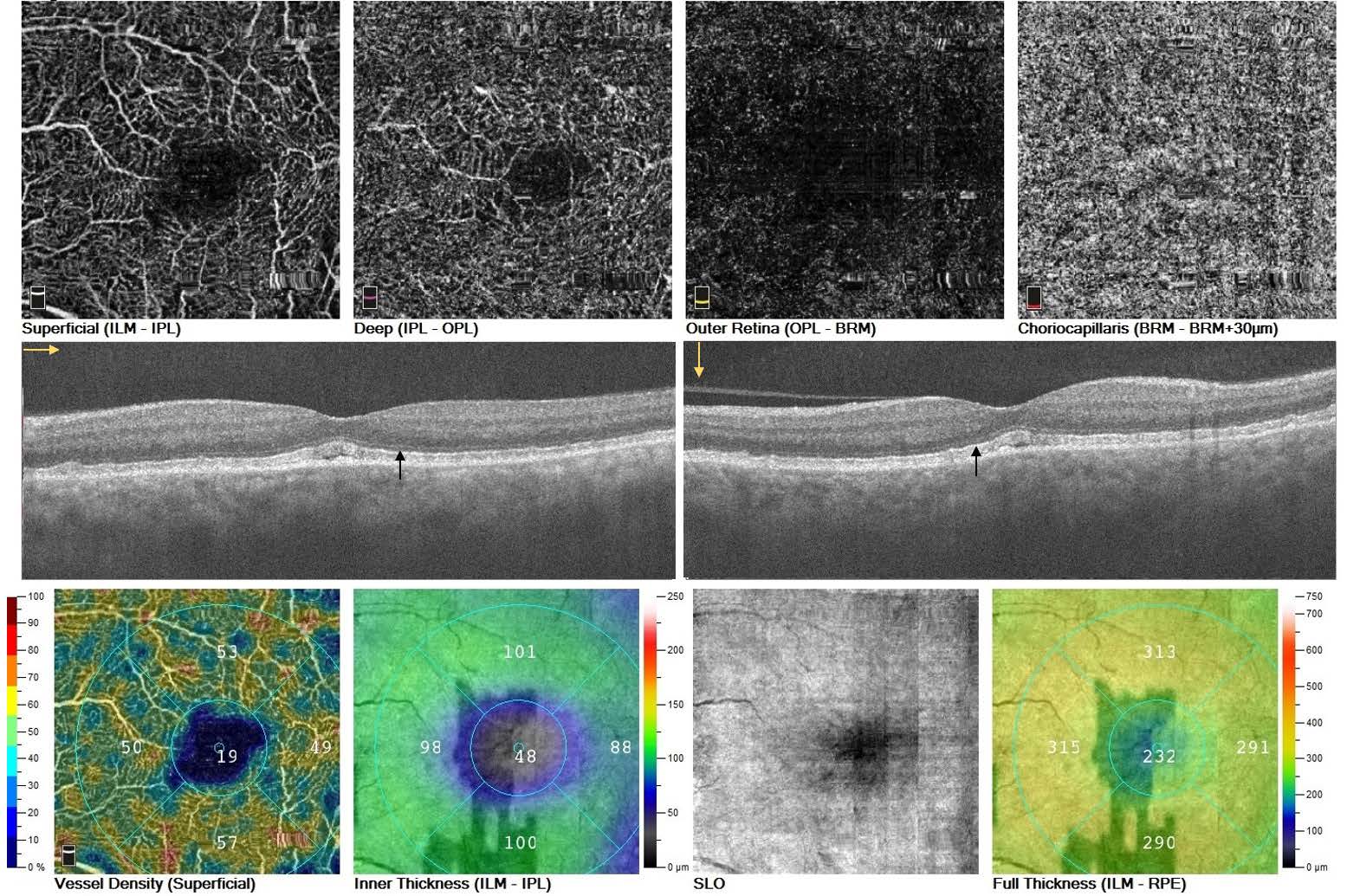
cystoid macular edema and subretinal fluid resolution. However, the outer segments showed irregularities and
thickening (black arrows). The en face images (top row) showed normal flow signals in the superficial, deep,
and outer retina. Note that some motion artifacts were present.
Discussion
COVID-19 is a systemic infection that can lead to life-threatening multi-organ involvement.3 The World Health Organization listed the BBIBP-CorV (Sinopharm) vaccine, an aluminum-hydroxide-adjuvanted inactivated whole virus vaccine, for emergency use on May 7, 2021, for adults 18 years and older. Two doses of the vaccine with a spacing of 3-4 weeks have an efficacy of 79% against symptomatic SARS-CoV-2 infection and hospitalization 2 weeks or more after the second dose.4 Rare ocular adverse events have been temporally associated with the vaccine. In a case series, Pichi et al. reported episcleritis (1 case), anterior scleritis (2 cases), acute macular neuroretinopathy (2 cases), paracentral acute middle maculopathy (1 case), and subretinal fluid (1 case) in 7 patients, which occurred within 2 weeks after receiving the Sinopharm COVID-19 vaccine.2 In our patient, unilateral serous macular detachment and CME were diagnosed 1 week after the second dose of the Sinopharm COVID-19 vaccination and resolved spontaneously and gradually within 10 days. Serous retinal detachment accompanying CME has been observed in diabetic macular edema, retinal vein occlusion, uveitis, chronic central serous chorioretinopathy, choroidal vascular lesions, intracameral cefuroxime toxicity, Coats' disease, Waldenström macroglobulinemia, and hypotony maculopathy.5-11 The authors did not observe any findings related to the these other conditions in the differential diagnoses.
Our patient was taking several medications, some of which have been linked to macular abnormalities. The results of an earlier case-control study indicated a potential association between 5-alpha reductase inhibitors, specifically finasteride, and the development of cystoid macular abnormalities and foveal cavitation, which may progress to the formation of a macular hole.12 A case report indicated that hydrochlorothiazide may be associated with CME. Notably, the CME observed in that case was bilateral and not accompanied by serous retinal detachment. Importantly, the CME resolved at 3 months with discontinuation of the medication, implying that it may not be relevant to the current case.13 It is well known that tight junctions between endothelial cells of retinal vessels and between retinal pigment epithelium form the inner blood-retinal barrier (BRB) and outer BRB, respectively. Damage to the inner BRB and subsequent increased permeability of the retinal capillary plexus may lead to macular edema. Furthermore, outer BRB disruption causes subretinal fluid accumulation. Cytokines and other pro-inflammatory mediators can lead to BRB dysfunction with or without structural vascular alterations. Degranulation of the mast cells, residing beside choroidal vessels, and the consequent release of histamine is another important cause of outer BRB alteration.14 Although vaccines containing live-attenuated viruses have the potential to transform to a wild type, especially in immunocompromised patients, most of the post-vaccination adverse events are immunemediated.
Microbial antigens or adjuvants (e.g., aluminum salts) and other additional components, present in vaccines, can trigger allergy and autoimmunity.15 Thus, in the patient reported here, an immune-mediated response triggered by the vaccine could have led to transient inflammation or allergic reaction, leading to inner and outer BRBs dysfunction and subsequent intraretinal and subretinal fluid accumulation. The resolution of the acute phase of inflammation and the clearance of inflammatory and allergic mediators may explain the spontaneous resolution of fluid accumulation. However, owing to the lack of fluorescein and indocyanine green angiography imaging and comorbidities in the patient, other causes cannot be definitively ruled out.
Conclusion
This report raises the possibility that Sinopharm COVID-19 vaccination may be associated with transient and self-limited serous macular detachment accompanying CME in some individuals. Additional work may further clarify any potential causative relationship and explore a plausible pathophysiological mechanism.
References
1. Ng XL, Betzler BK, Testi I, et al. Ocular Adverse Events after Covid-19 Vaccination. Ocul Immunol Inflamm. 2021;29:1216-1224.
2. Pichi F, Aljneibi S, Neri P, et al. Association of Ocular Adverse Events with Inactivated Covid-19 Vaccination in Patients in Abu Dhabi. JAMA Ophthalmol. 2021;139:1131-1135.
3. Rello J, Belliato M, Dimopoulos MA, et al. Update on Covid-19 in the Intensive Care Unit from the 2020 Hellenic Athens International Symposium. Anaesth Crit Care Pain Med. 2020;39:723-730.
4. World Health Organization. Interim recommendations for use of the inactivated COVID-19 vaccine BIBP developed by China National Biotec Group (CNBG), Sinopharm. World Health Organization; 2022. https://www.who.int/publications/i/item/WHO-2019-nCoV-vaccines-SAGE-rec…
5. Accorinti M, Okada AA, Smith JR, et al. Epidemiology of Macular Edema in Uveitis. Ocul Immunol Inflamm. 2019;27:169-180.
6. Baker PS, Garg SJ, Fineman MS, et al. Serous Macular Detachment in WaldenströmMacroglobulinemia: A Report of Four Cases. Am J Ophthalmol. 2013;155:448-455.6
7. Chlasta-Twardzik E, Nowińska A, Wylęgała E. Acute Macular Edema and Serous Detachment on the First Day after Phacoemulsification Surgery: A Case rRport. Am J Ophthalmol. 2020;20:100905.
8. Kokame GT, de Leon MD, Tanji T. Serous Retinal Detachment and Cystoid Macular Edema in Hypotony Maculopathy. Am J Ophthalmol. 2001;131:384-386.
9. Muraoka Y, Murakami T, Nishijima K, et al. Association Between Retinal Venular Dilation and Serous Retinal Detachment in Diabetic Macular Edema. Retina. 2014;34:725-731.
10. Otani T, Yamaguchi Y, Kishi S. Serous Macular Detachment Secondary to Distant Retinal Vascular Disorders. Retina. 2004;24:758-762.
11. Van Rijssen TJ, Van Dijk EHC, Yzer S, et al. Central Serous Chorioretinopathy: Towards an Evidence-Based Treatment Guideline. Prog Retin Eye Res. 2019;73:100770.
12. Shin YK, Lee GW, Kang SW, et al. Macular Abnormalities Associated with 5α-Reductase Inhibitor. JAMA Ophthalmol. 2020;138:732-739.
13. Iacono P, Battaglia PM, Bandello F. Macular Edema Associated with Hydrochlorothiazide Therapy. Br J Ophthalmol. 2011;95:1025, 1036-1027.
14. Daruich A, Matet A, Moulin A, et al. Mechanisms of Macular Edema: Beyond the Surface. Prog Retin Eye Res. 2018;63:20-68.
15. Cheng JY, Margo CE. Ocular Adverse Events Following Vaccination: Overview and Update. Surv Ophthalmol. 2022;67:293-306.
Statement of Ethics
This report followed the tenets of the Declaration of Helsinki and was approved by the Institutional Ethics Committee (IR.SEMUMS.REC.1400.167, Research Ethics Committee of Semnan University of Medical Sciences and Health Services). Written informed consent has been obtained from the patient for the publication of this case report.
Conflict of Interest Statement
The authors declare no conflicts of interest related to this topic.
Funding
This work received no funding or grant support.
Authorship
We attest that all authors contributed significantly to the creation of this manuscript, each having fulfilled the criteria, as established by the ICMJE.