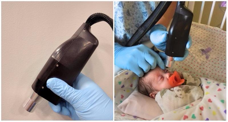
Researchers pioneer handheld, 3D, and inter-surgical use of optical coherence tomography in pediatric settings.
In the early 1990s, a revolutionary new technology began to emerge in the field of ophthalmology. It was called optical coherence tomography (OCT), and it represented the first major advance in retinal imaging for almost 150 years.
OCT uses light waves to create a cross-sectional image of the retina—similar to the way sound waves are used in a prenatal ultrasound—allowing physicians to see beneath the surface of the retina for the first time. When operated by a trained technician in a clinic, OCT overcomes one of the major limitations of earlier devices such as the ophthalmoscope, which revealed only a surface view of the retina.
“Using an ophthalmoscope is like looking at the ocean from above,” explains Dr. Cynthia Toth, professor of ophthalmology in the Duke University School of Medicine. “You see the beautiful pattern of waves on top, but there’s a lot going on below the surface, too, and it can be really important to see that.”
Toth, who is a practicing retinal surgeon, and her longtime collaborator, Dr. Joseph Izatt, have been at the forefront of OCT innovation virtually since its inception, helping it grow from an idea to the standard of care for adult patients.
Now, with support from the Duke Clinical and Translational Science Institute (CTSI), they are developing a suite of devices and techniques that take OCT out of specialized clinics, allowing it to be be used at a patient’s bedside—or even in surgery—to give physicians an unprecedented level of real-time information.
Window to the brain
“What we at Duke are known for in the OCT world is pioneering the use of handheld probes for pediatric imaging,” says Izatt, the Michael J. Fitzpatrick Professor of Engineering at Duke.
In 2012, a handheld device developed by Toth and Izatt with partial funding from Duke’s Clinical and Translational Science Award was approved by the FDA for use in humans. Izatt took the device to market via a startup called Bioptigen, and for the first time, there was a commercial alternative to the conventional OCT system—a bulky tabletop setup that is typically located in an eye clinic’s photography suite and operated by a technician.
While the conventional system requires that a patient be able to follow instructions, such as opening and closing their eyes, looking in different directions, and keeping still, Toth and Izatt’s handheld probe can be used to examine infants and other patients who are unable to respond to instructions. That probe is now used worldwide in nurseries and under anesthesia, allowing physicians to get cross-sectional images of a retina in real time. This can provide them with critical, time-sensitive health information that goes beyond the eyes themselves.
“The retina is an extension of the brain, so it’s a window in,” says Toth. OCT images can reveal neurological markers that correspond to conditions affecting the brain, and help physicians assess whether a treatment is working.
One such condition is hypoxic ischemic encephalopathy (HIE), a brain injury resulting from a lack of oxygen. HIE is rare, but serious—resulting in impairment or death in more than half of all cases. To avert the worst effects, early diagnosis is key.
“If you recognize the condition in the first six hours, you can intervene,” Toth says. “But you have to identify it early and fast. You can do an ultrasound or brain MRI, but a hospital typically won’t do that until five or ten days after birth.”
Portable OCT could offer a fast-acting way to examine the neuromarkers of an infant suspected of having HIE. Toth and Izatt have published findings supporting the idea that hypoxic injury to the brain also affects retina thickness, a link that had not been investigated before. As they continue to gather data, early results indicate that diagnosing HIE based on retinal imaging may be possible.
Going 3D
Five years after their handheld probe was approved for use in humans, Toth and Izatt have continued to expand the frontiers of OCT.
In 2016, with support from Duke CTSI, they began testing an improved probe that puts powerful new functionality into a more compact, easy-to-use form factor.
“The previous commercial device only does cross-sections. What CTSI funded is a new device that gets full-volume, three-dimensional images,” Izatt says.
About the size of an umbrella handle, Toth says, “It’s smaller, it’s lighter, and it’s faster. So I can do non-dilated examination and get images through the pupil. One of the things we watch is how the pupils respond, so I can’t use the dilating drops. This system was designed to allow us to do that.”
The ability to do 3D imaging also opens OCT to a new arena—the operating room. “My dream has been to be able to use OCT pictures in surgery,” Toth says.
With 3D, a surgeon isn’t restricted to a surface or cross-sectional view—they can adjust the image in any direction to interactively monitor the retina as they operate. Toth explains: “It becomes image-guided surgery, and you’re actually watching it dynamically as it’s changing. With OCT I can see and respond as the tissue moves.”
Toth and Izatt’s inter-surgical OCT research system at Duke is currently the only one in the world. They are continuing to hone the technology and establish best practices for its use. Ultimately, they envision making the full suite of OCT tools more user friendly.
“These are pretty complicated instruments to use,” Izatt says. “There are a lot of parameters that have to be controlled simultaneously while attending to a baby that may be crying or struggling. We’re interested in ease of use for these devices, with features like auto-focus, and in making them smaller and lighter.”
Toth adds, “I’m lucky to have the best OCT handheld imagers in the world in my lab. But we want any care provider to be able to do this, and do it well.”
In the meantime, they are continuing to search for the next revolutionary idea.
"I’m thrilled, because OCT is taking off, and federal grant dollars have made a real impact,” Toth says. “Working at a university and with CTSI is so exciting, because we can brainstorm with smart people across the university and come up with what we think we should be doing next. I don’t want to just focus on one idea or technology—I like looking forward to the next idea and the next technology.”
This research was supported in part by Duke's Clinical and Translational Science Award, NIH grant number UL1TR001117.
This story was originally published on the Duke CTSI website.