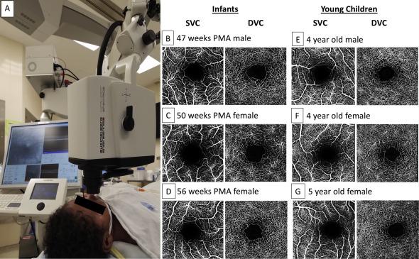
A portable optical coherence tomography angiography (OCT-A) system can be used for depth-resolved visualization of retinal microvasculature in infants, according to a study published in Ophthalmology Retina in 2018. The study is the first to image the superficial and deep parafoveal retinal microvasculature in healthy, full-term infants.
Previously, visualization of the perifoveal microvasculature of healthy infants was limited to histopathological studies, and earlier studies did not examine the superficial and deep vascular complex.
The Duke researchers also showed that the retinal vasculature of infants 47 to 56 weeks postmenstrual age (PMA) is similar to that of young children and adults with healthy retinas, suggesting that the parafoveal microvasculature in both the superficial and deep vascular complexes is formed by 47 weeks PMA. The results are in line with previous histologic studies.
Imaging with portable OCT-A has utility beyond studying infant retinal vascular development, says the study’s senior author, Lejla Vajzovic, MD, a pediatric retinal specialist at Duke.
Vajzovic expects the new system to have potential uses in diagnosis and disease monitoring, guiding the way for progress in pediatric ophthalmology.