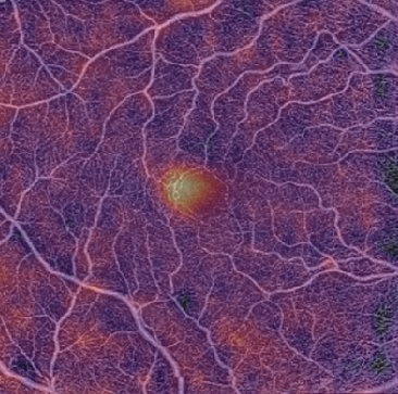
 Duke Reading Center is a comprehensive image reading center that specializes in reliable, timely, reproducible and systematic analysis of images captured by many different retinal imaging technologies, including optical coherence tomography, fundus autofluorescence, color fundus photography and fluorescein angiography. The Duke Reading Center is dedicated to quality, efficiency and innovation by constantly improving and refining research protocols and embracing cutting-edge technology and research techniques.
The Duke Reading Center has been operational since the introduction of OCT into multicenter clinical trials in 2001. Our experienced staff is capable of handling trials, of any study size or duration, involving many different treatments for various ophthalmic diseases. Since its inception, the Reading Center has been involved in over 60 industry and NIH-sponsored multicenter clinical trials conducted in the US, Canada, Australia, South America, Europe and Asia. Our readers have provided image analysis for studies targeting a variety of ophthalmic diseases, including, but not limited to, diabetic macular edema, neovascular and non neovascular AMD, vitreoretinal interface disorders, neuro-ophthalmological diseases, uveitis and glaucoma. In addition, we have graded ocular images for safety studies of drugs used to treat non-opthalmological diseases, such as cardiovascular and psychiatric conditions. These studies have ranged in size from 20 to 2500 subjects, with 3-24 separate image sets collected per subject. Our readers have also certified over 10,000 technicians in the capture and submission of ophthalmic images to the Duke Reading Center according to study-specific protocols.
Founded primarily as a contract research center, the Duke Reading Center advises sponsors about the different types of retinal imaging and how to optimally utilize them in a clinical trial setting. The Reading Center also receives de-identified images and provides analyses of these images according to study-specific protocols tailored to the conditions of each individual study. In addition, the Reading Center compiles images and performs statistical analysis of the image data. Contract research organizations, study sponsors and coordinating centers use this retinal imaging data to assess natural disease history and outcomes of interventional trials. The Reading Center also certifies, trains, and supports study-site personnel in hardware and software capabilities and maintains a record of all certifications granted to site technicians.
The Duke Reading Center comprises a group of ocular imaging, clinical trial and biostatistical specialists who are dedicated to delivering reliable and reproducible results quickly and accurately. Staff members known as readers analyze images and possess a wealth of experience that enables them to interpret images for multiple qualitative and quantitative morphological parameters. The Reading Center works closely with the Duke Clinical Research Institute, the largest academic contract research organization (CRO) in the world. We have partnered with the DCRI to build the Reading Center’s Data Transmission System (DTS), a powerful custom-designed web based tool that transmits images quickly and securely. DTS is fully CFR part 11 compliant; it requires password access, automatically monitors any changes to data and creates an internal audit trail. The Reading Center has handled studies utilizing numerous retinal-imaging modalities and has well-established, clear and efficient grading protocols that have been constantly refined over the past decade. The Reading Center continuously develops innovative grading techniques to incorporate the latest breakthroughs in retinal-imaging technologies into our work. Through our extensive quality assurance activities, we constantly strive to improve our research protocols, grading techniques, and intra- and inter-reader reproducibility. Our experience allows us to tailor our research protocols to meet the needs of nearly any trial involving ocular pathology and imaging, while delivering accurate, reproducible results in an expedient manner.
Director
Directors of Grading
Biostatistician
Administration
Dawn Cronce
Administrative Manager
Ellen Young
Operations and Invoice Manager
Kelly Inman
Senior Grants and Contracts Manager
Contact
Email: octrdctr@mc.duke.edu


