

Confocal imaging showing the interaction of nuclear LC3 (green) interacts with PML nuclear bodies (red) in trabecular meshwork cells treated with leptomycin B (Shim et al, Autophagy 2020).

Interactive 3D surface plot analysis visualizing the co-localization of ATG16L (green) at the basal body of the primary cilium upon cyclic mechanical stretch in trabecular meshwork cells. Primary cilia were stained using acetylated TUBA4A (Shim et al. PNAS, 2021).

Transmission electron microscopy images of cross-sectional areas of the myelinated segment of the optic nerve of DBA/2J::GFP-LC3, showing the presence of large autophagic vacuoles in the degenerating axons (Hirt et al. Cell Death Dis, 2018).
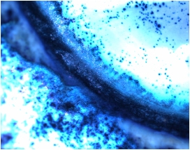
Human perfused anterior segment transduced with a recombinant adenovirus containing the reporter gene Lac Z.

Electron micrograph showing the intralysosomal accumulation of amorphous electron dense material within secondary lysosomes in oxidatively stressed trabecular meshwork cells.

Confocal microcopy image of trabecular meshwork cells stained with the acidotropic dye Lysotraker Red.

SA-b-gal staining in the glaucomatous outflow pathway.

Histological section of human trabecular meshwork transduced with an adenovirus containing the reporter gene LacZ under the Chitinase 3-like 1 promoter region.