When it is time to take the reins as the primary surgeon for your first cataract surgery, you must be prepared with a strong foundation of knowledge. Here we outline four basic yet important pearls that will prepare you and help you excel — all while reducing your stress level.
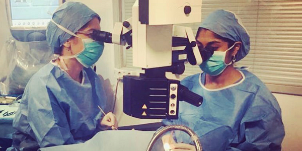
1. Know your microscope. Understanding the settings of your operating microscope is one of the most fundamental steps of cataract surgery. Take a day out of your busy schedule to learn the basics of your microscope with a senior resident, fellow or attending. Know how to turn on the microscope. Determine if you will be operating from a temporal or superior approach and ensure that you orient the oculars in the right position for yourself and your assistant.
Identify the center focus/XY button and remember to press it at the beginning of a new case so that your microscope is reset before surgery begins. Check your interpupillary distance and then zero both objectives such that you are not seeing double images or overaccommodating through an incorrect refractive error. Practice being able to move the microscope manually using the microscope handles. All of these skills, although seemingly simple, are critical in keeping a smooth case flow. The last thing you want to do is fumble with the microscope before your first case and feel embarrassed!
2. Know your foot pedals. During cataract surgery, all four of your extremities will be constantly active. Not only will you be focused on the fine and precise movements of both your hands throughout the case, but you will also need to use both of your feet simultaneously to control the pedals of your microscope and phacoemulsification machine.
Let’s first focus on the microscope foot pedal. Most cataract surgeons will use their nondominant foot for the microscope foot pedal and their dominant foot for the phaco pedal; however, this may vary based upon your training institution and personal preferences. Many ophthalmologists prefer to remove their shoes so that they can better feel the foot pedals. I actually operate with thin-soled shoes that help me feel the pedals and preclude the need to remove my shoes.
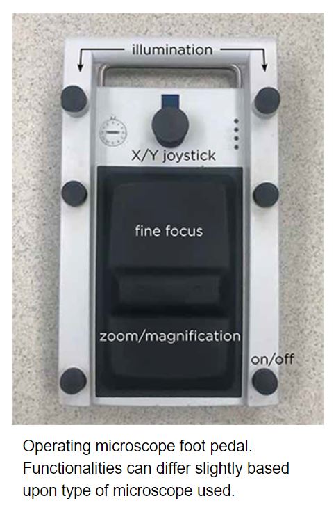
What are the main functions to know? There is an on/off button on the lower aspect of the pedal. The joystick on the upper aspect allows you to control the XY position of the scope. There is a lower switch (which is controlled by the heel of your foot) to modulate the zoom and magnification of the microscope, and there is an upper switch (which is controlled with the ball of your foot) that will raise and lower the microscope and make small focus changes.
You should manually move the microscope with the handles to center the surgical eye and achieve gross focus prior to starting surgery so that you can ensure maximal excursion of the XY joystick as well as the zoom, magnification and focus functions. There are also switches above the joystick that you can use to increase and decrease illumination. Throughout the procedure, you need to dynamically change the lighting, magnification and focus in order to improve your visualization and surgical precision.
The phaco pedal has its own unique features, and there are three main positions: position 1 is irrigation, position 2 is both irrigation and aspiration and position 3 is irrigation, aspiration and ultrasound power. Rapid and accurate foot movements are necessary to adjust fluidics and power throughout the procedure, and this takes practice to master. Aspiration in position 2 is controlled in a linear fashion: as you depress the foot pedal further, more aspiration is achieved.
Ultrasound power is similarly controlled in position 3. In addition, you can activate different ultrasound modes, such as pulse and burst modes, in this position to help emulsify the lens nucleus. Depending on the surgical step you are performing, you can also use the switches on the foot pedal to move between modes on the phaco machine (including sculpt, chop, quadrant, epinucleus, irrigation and aspiration, polish and anterior vitrectomy). I would learn to independently move through these modes with your foot pedal so that you do not have to depend on your scrub nurse to change settings for you in the midst of a critical step of the surgery.

3. Know your phacoemulsification machine and phacodynamics. There are four main phacodynamic factors that all beginning surgeons need to know.
-
Power is the ultrasound energy used to break up the cataract.
-
The aspiration rate is the rate at which fluid enters the phacoemulsification tip, which can, in turn, control the speed with which pieces of lens come to the tip. A higher aspiration rate can be advantageous for a faster, more experienced surgeon, while a lower aspiration may be beneficial for beginning surgeons — or in scenarios such as a posterior capsular rupture.
-
Vacuum is defined by how strongly the nucleus pieces are held by the phaco tip. Higher vacuum settings are often required during chopping maneuvers in order to establish strong purchase of the lens nucleus.
-
Many phaco machines rely on bottle height, where the inflow of fluid into the eye is based on gravity fluidics. New-generation machines now use infusion pumps to maintain a chosen intraocular pressure in the eye and ensure stability of the anterior chamber.
These four factors are modulated based upon which step of cataract surgery you are performing. Your phaco machine alters these factors in its different modes, and you as the surgeon can titrate settings as needed during different points of the procedure to optimize your performance. Consider taking the time to sit down with a manufacturer’s representative of the platform you will be using during your training, and make sure to customize the settings as you perform more cases and develop new preferences. These reps are also invaluable when it comes to troubleshooting any unexpected errors that can occur during surgery.
4. Know your patient and the instruments you need. Even before entering the operating room, it is critical to know your patient, their ocular history, the type of cataract they have and their refractive goals. Pay careful attention to identify any factors that could render your surgery more challenging, such as poor dilation, shallow anterior chamber, pseudoexfoliation, phacodenesis or prior ocular trauma or retina surgery. You must have a plan of attack if you identify any of these.
In addition, you should select a primary intraocular lens (IOL) to place in the capsular bag as well as backup lenses (such as a sulcus lens or anterior chamber lens) in the event there is a posterior capsular rupture. It is important to review how to calculate the power of these various types of lenses.
Prior to surgery, you should also mentally rehearse the steps of cataract surgery and the instruments you will use. Imagine where your paracentesis would be created, which type of viscoelastic would be injected, where the main wound would be created, what maneuvers you would use to create a circular capsulorhexis in case it begins to radialize, how to perform careful hydrodissection and hydrodelineation, what method of nuclear disassembly you will use and, lastly, how you will remove cortical material and inject the IOL implant.
It takes years for these seemingly straightforward steps to become second nature, but you will not regret it.