This case originally appeared in Duke Heath's Clinical Practice Today monthly ophthlalmology newsletter.
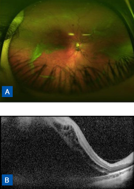
After presenting with blurry vision and reporting the presence of dark spots in her vision, an 8-year-old girl was diagnosed with bilateral retinal detachment and referred to the Duke Pediatric Retina and Optic Nerve Center at the Duke Eye Center (Figure 1). She had a family history of retinal detachment, and her father had type 1 Stickler syndrome.
FIGURE 1 to the right. (A) Fundus photography of right eye indicating prominent vitreous veils and retinal detachment. (B) Optical coherence tomography shows subretinal fluid extending into macula.
Question: What steps needed to be taken for this patient in addition to repairing her retinal detachments?