Diabetic eye disease is a prime target for remote diagnosis and management.
Ophthalmology has been one of the hardest-hit specialties during the coronavirus pandemic.1 Although medicine has significantly increased video and telephone visits during these precarious times,2,3 the question is how much ophthalmology, and in particular the retina subspecialty, can rely on telemedicine. Unfortunately, for most ophthalmologists, there is no easy access to remote imaging technology or screening tools. The pandemic has spurred some retinal specialists to develop innovative techniques for do-it-yourself at-home retinal imaging.4 Although this may not be practical for all patients and practices, it underscores a critical need to expand access to remote care in ophthalmology.
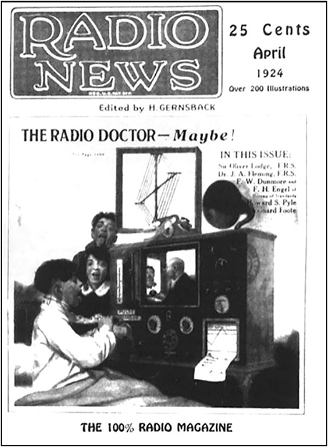
The first available records of telehealth date to 1878 when The Lancet published a letter to the editor describing how the newly invented telephone could improve medical diagnoses via studying the sound produced by a muscle.5 The same year, another article in Popular Science Review postulated that the telephone might be used in auscultation of heart and lung sounds.5 In April 1924, Radio News magazine published an innovative cover story: “The Radio Doctor – Maybe!” in which the “radio doctor” was linked to a patient not only by a sound but also by a live picture (Figure 1); this was forward-thinking, as the radio had just begun to reach American homes, and the first video transmission would come years later.6 In 1925, Hugo Gernsback, a pioneer in radio technology, published in Science and Invention his ideas for the Teledactyl, an integrated video system with a sensory feedback device that would allow a physician to interview and examine a patient from afar.7 Those early fantasies moved into a real telemedicine context for the first time in 1948 when radiologic images were transmitted by telephone between West Chester and Philadelphia (a 24-mile distance).8 The first anecdotal records of teleophthalmology started with the Apollo missions and retinal circulation monitoring. Over the years, tele-eye care evolved to the broader use of different methodologies assessing mostly glaucoma, retinopathy of prematurity (ROP), and diabetic retinopathy (DR). Currently, there are 2 telemedicine approaches available in ophthalmology: the “store and forward” method (asynchronous electronic transmission of health data), and live audio-video telemedicine (2-way synchronous live communication between patient and provider).9

Figure 1. April 1924 Radio News cover depicting a “radio doctor;” then a fantasy, and now a reality.
The COVID-19 reality is accelerating an anticipated need for increased teleophthalmology presence. In December 2016, the US Health Resources and Services Administration predicted a shortage of more than 6,000 ophthalmologists by 2025.10 Additionally, many patients from rural or remote locations have very limited access to eye care. Teleophthalmology provides an avenue for expanding access to care in remote geographical areas, as well as for patients with mobility, health, and transportation concerns. Furthermore, screening eye examinations are becoming increasingly mandated in the United States due to the Affordable Care Act.11 Traditional screening for eye disease typically requires examinations by eye care specialists through dilated pupils. However, only a fraction of patients at risk receive such screening. Teleophthalmic examination is associated with high effectiveness and may be an alternative to live examination. Despite this tremendous promise, several barriers, especially the access, cost, reimbursement, and lack of universally accepted protocol, prevent it from being widely accepted in the United States.
TELEOPHTHALMOLOGY IN RETINAL DISEASE
Multiple retinal diseases can be successfully assessed using the tele-eye care approach, including DR, ROP, and age-related macular degeneration (AMD). Several characteristics make DR ideal for this methodology: it poses a significant health risk in the population, it has identifiable presymptomatic stages, it can be easily detected by retinal imaging, and there are early treatments available to prevent vision loss.12 In the United States, DR is the leading cause of preventable vision loss in work-age adults.13,14 Although early diagnosis and timely treatment can decrease the risk of severe vision loss by more than 90%, it is estimated that only 40% to 50% of patients are adherent to the recommended screening intervals.15,16 Even for patients in urban settings with health insurance and access to care, the minimum recommendations for DR screening are frequently not met due to difficulty scheduling appointments and excessive wait times.13
The American Academy of Ophthalmology publishes preferred practice patterns for DR screening every few years.17 Patients with type 1 diabetes with onset before age 30 should have their first screening exam 5 years after diagnosis and every year after that. Patients with late-onset type 1 diabetes or a new diagnosis of type 2 diabetes should undergo a dilated retinal examination at the time of diagnosis and yearly after that. Irrespective of clear guidance, patients’ adherence to the screening and follow-up is suboptimal, resulting in poor visual outcomes.15,16
TELEOPHTHALMOLOGY IN DIABETIC RETINOPATHY
Because they have multiple comorbidities, diabetic patients may be the most compliant with their primary care provider (PCP) follow-ups, making primary care clinics an ideal location to implement retinal image capture for DR screening. Retina screening in this setting has multiple advantages: accessibility, patient capture, cost-effectiveness, improved patients triage (eg, advanced stages of disease that need retina treatment get scheduled to retina clinics, while early stages go to comprehensive ophthalmology or optometry).
Teleophthalmology has been shown to increase compliance with the proposed guidelines and decrease the number of patients who miss recommended screenings;18 however, it is seldom used in the United States. Current teleophthalmology initiatives in the United States include the Veterans Health Administration DR screening program, the Indian Health Service-Joslin Vision Network DR screening program, and a few pilot studies in emergency departments and primary care clinics.19,20 There is no widespread and uniformly accepted DR screening modality currently available outside these institutional networks.
A NEW DIABETIC RETINOPATHY SCREENING INITIATIVE
Duke Ophthalmology recently launched a DR telehealth initiative with a few simple goals: improved access, early diagnosis (thus, improving visual outcomes), and reduced health care costs. The general idea has been to place simple-to-use nonmydriatic color fundus and optical coherence tomography (OCT) cameras in Duke primary care clinics to screen every diabetic patient on the way out. Although initially located at the PCP clinics, the ultimate goal has been a kiosk-based screening model.
The pilot program involved remote diagnosis that was separate from traditional teleophthalmology in several key ways. The conventional method takes color fundus photography of dilated pupils and is performed by expert photographers with expensive equipment in a dedicated brick-and-mortar imaging center. Remote diagnosis is the use of color fundus photography and OCT on nondilated pupils and is performed by nonexpert imagers using less expensive equipment permanently located at the point of service (eg, primary care clinic). This approach improved patient convenience and capture.21 Moreover, it significantly increased the Healthcare Effectiveness Data and Information Set (HEDIS) measures, which turned out to be an excellent incentive for participating clinics.
BARRIERS TO IMPLEMENTATION
Regardless of the value of teleophthalmology, there are many limitations to its widespread adoption. These include logistical complexity, lack of protocol consensus in imaging, financial model, and reimbursement issues, as well as the liability associated with performing remote examinations and assessments. Significant efforts have been made to establish universal screening protocols, but none yet exist regarding best imaging modality, minimum necessary image quality, or grading rubric.
Individual screening centers can currently select from a variety of imaging modalities. Options for screening patients include a single color fundus photo, a 7-field Early Treatment of Diabetic Retinopathy Study (ETDRS) standard compilation, an OCT image, or an ultrawidefield Optos image. There is no consensus about the most cost-effective screening tool, nor has the best screening tool for optimal sensitivity and specificity been recently addressed. Furthermore, assuming the images are clear and appropriate, they must be graded for severity. The ETDRS is the most commonly used metric for classifying DR severity.22 To simplify the classification, physicians can also use the recently proposed International Clinical Diabetic Retinopathy Disease Severity Scale, based on ETDRS and the Wisconsin Epidemiologic Study of Diabetic Retinopathy.23 In addition, there is little regulatory guidance and no required standard for image quality. In general, the lack of standardization can compromise the quality of care provided by telescreenings and undermine its utility.
Also, the liability associated with teleretinal screening provides a substantial barrier to its expansion. Image misreading or failing to promptly refer a patient can result in irreversible visual repercussions. In addition, a secure electronic transfer of patient information and images from the site of collection to the site of interpretation must be provided to protect patients’ rights.
INCORPORATING NEW TECHNOLOGIES INTO TELEOPHTHALMOLOGY FOR DR
Current events have prompted a significant change in health care delivery in general and encouraged the use of video and telephone visits. As attractive as this approach is, the question remains about how useful it is to retina specialists without retinal imaging. As discussed above, diagnosis of retinal pathologies heavily relies on seeing (eg, retinal exam and imaging), and while telemedicine in the form of video and telephone visits carries the same liability as in-person examination, without the supporting imagining, video or telephone visits have limited merit to the retinal subspecialty. Nonetheless, they can successfully be used in the form of video triage or as a follow-up consultation for the study-only visit. Additionally, advancements in imaging technologies and automated image interpretation will lead to overcoming these shortcomings and delivering on the promises of teleophthalmology.
For instance, over the past few years, there has been increased use of deep learning to identify and classify DR successfully; thus, automating and improving the screening process reliability.24,25 This approach can eliminate concerns related to standardized interpretation and mitigate the liability currently shouldered by the reading physician. Ideally, a deep-learning algorithm would be integrated into the imaging machine’s software at the remote location, allowing for a fast diagnosis of retinal pathology and preferably automatically referring to an eye specialist. Despite the significant advances, clearly, much work is ahead of us.
CONCLUSION
The coronavirus pandemic has highlighted a clear need for a platform to assist with patients’ care without physical contact, while still providing excellent service and security for both patients and physicians. To achieve the best possible care and improve visual outcomes, the retina community must develop standardized methods for retinal imaging and interpretation. A number of noninferiority studies involving diverse imaging modalities lay ahead of us. We also have to assess further the cost, visual outcomes, and patients’ satisfaction in these settings. Finally, we should strengthen multidisciplinary collaboration focused on the development of deep-learning methods for high-accuracy image interpretation as well as less expensive and more portable (kiosk or home-based) imaging devices that are accepted by the retina community.
REFERENCES
- Mehrotra A, Chernew M, Linetsky D, Hatch H, Cutler D. The Impact of the COVID-19 pandemic on outpatient visits: a rebound emerges. May 19, 2020. https://www.commonwealthfund.org/publications/2020/apr/impact-covid-19-…;
- Mehrotra A, Ray K, Brockmeyer DM, Barnett ML, Bender JA. Rapidly converting to “virtual practices”: outpatient care in the era of Covid-19. NEJM Catalyst Innov Care Deliv. 2020;1(2).
- Wosik J, Fudim M, Cameron B, et al. Telehealth transformation: COVID-19 and the rise of virtual care. J Am Med Inform Assoc. 2020;27(6):957-962.
- Salzman IJ. A retina telemedicine technique. May 2020. Accessed August 12, 2020. https://www.aao.org/eyenet/article/a-retina-telemedicine-technique
- Aronson SH. The Lancet on the telephone 1876-1975. Med Hist. 1977;21(1):69-87.
- Field MJ, ed. Telemedicine: A Guide to Assessing Telecommunications for Health Care. National Academies Press; 1996.
- Novak M. Telemedicine predicted in 1925. Smithsonian Magazine. 2012, March 14. Accessed August 24, 2020. https://www.smithsonianmag.com/history/telemedicine-predicted-in-1925-1…;
- Gershon-Cohen J, Cooley AG. Telognosis. Radiology. 1950;55(4):582-587.
- Gioia G, Salducci M. Medical and legal aspects of telemedicine in ophthalmology. Rom J Ophthalmol. 2019;63(3):197-207.
- US Department of Health and Human Services. National and regional projections of supply and demand for surgical specialty practitioners: 2013-2025. Author; Rockville, MD; 2016. https://bhw.hrsa.gov/sites/default/files/bhw/health-workforce-analysis/…;
- Wong TY, Sun J, Kawasaki R, et al. Guidelines on diabetic eye care: the International Council of Ophthalmology recommendations for screening, follow-up, referral, and treatment based on resource settings. Ophthalmology. 2018;125(10):1608-1622.
- Surendran TS, Raman R. Teleophthalmology in diabetic retinopathy. J Diabetes Sci Technol. 2014;8(2):262-266.
- Salongcay RP, Silva PS. The role of teleophthalmology in the management of diabetic retinopathy. Asia Pac J Ophthalmol. 2018;7(1):17-21.
- Cheung N, Mitchell P, Wong TY. Diabetic retinopathy. Lancet. 2010;376(9735):124-136.
- Liu Y, Zupan NJ, Shiyanbola OO, et al. Factors influencing patient adherence with diabetic eye screening in rural communities: a qualitative study. PLoS One. 2018;13(11):e0206742.
- Murchison AP, Hark L, Pizzi LT, et al. Non-adherence to eye care in people with diabetes. BMJ Open Diabetes Res Care. 2017;5(1):e000333.
- Flaxel CJ, Adelman RA, Bailey ST, et al. Diabetic retinopathy preferred practice pattern. Ophthalmology. 2020;127(1):P66-P145.
- Conlin PR, Fisch BM, Cavallerano AA, Cavallerano JD, Bursell SE, Aiello LM. Nonmydriatic teleretinal imaging improves adherence to annual eye examinations in patients with diabetes. J Rehabil Res Dev. 2006;43(6):733-740.
- Rathi S, Tsui E, Mehta N, Zahid S, Schuman JS. The current state of teleophthalmology in the United States. Ophthalmology. 2017;124(12):1729-1734.
- Legarreta JE, Conner IP, Loewen NA, Miller KV, Wingard J. The utility of iPhone-based imaging for tele-ophthalmology in a triage capacity for emergency room consultations. Invest Ophthalmol Vis Sci. 2014;55(13):4876-4876.
- Hadziahmetovic M, Nicholas P, Jindal S, Mettu PS, Cousins SW. Evaluation of a remote diagnosis imaging model vs dilated eye examination in referable macular degeneration. JAMA Ophthalmol. 2019;137(7):802-808.
- Early Treatment Diabetic Retinopathy Study Research Group. Fundus photographic risk factors for progression of diabetic retinopathy. ETDRS report number 12. Ophthalmology. 1991;98(5 Suppl):823-833.
- Wu L, Fernandez-Loaiza P, Sauma J, Hernandez-Bogantes E, Masis M. Classification of diabetic retinopathy and diabetic macular edema. World J Diabetes. 2013;4(6):290-294.
- Korot E, Wood E, Weiner A, Sim DA, Trese M. A renaissance of teleophthalmology through artificial intelligence. Eye. 2019;33(6):861-863.
- Walton OBt, Garoon RB, Weng CY, et al. Evaluation of automated teleretinal screening program for diabetic retinopathy. JAMA Ophthalmol. 2016;134(2):204-209.