Duke has a long history of pioneering Optical Coherence Tomography (OCT) technology that has revolutionized patient care for adults and pediatric patients.
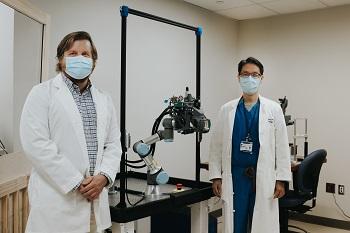
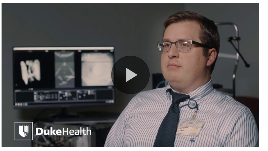
Duke clinician scientist Anthony Kuo, MD, Associate Professor of Ophthalmology and corneal specialist; and research scientist Ryan McNabb, PhD have developed a robotically aligning OCT system that captures a three-dimensional image of the entire eye, all while allowing the system operator to maintain a safe physical distance from the patient. This robotically aligning OCT system builds off of a predecessor prototype system developed by Pratt School of Engineering collaborators Mark Draelos, MD, PhD, Pablo Ortiz, and Joseph Izatt, PhD.
“This is the first, to our knowledge, robotically aligning OCT system designed for potential clinical use in both normal patients and those with pathologies. This system offers the ability to safely distance operator and patient while providing the additional imaging benefits provided by active tracking and compensation,” said Kuo.
A Pioneer in OCT
The work of Kuo and McNabb extend Duke’s position as a pioneer in OCT technology, which began with the work of Cynthia Toth, MD, Joseph A.C. Wadsworth Distinguished Professor of Ophthalmology, and Joseph Izatt, PhD Michael J. Fitzpatrick Distinguished Professor of Engineering, both having dual appointments in each respective department. Toth and Izatt have developed applications of intraoperative OCT that allows more precise ophthalmic surgery and a handheld OCT that is particularly useful imaging infants in the NICU and pediatric patients.
“We are proud to continue the well-recognized contributions of Duke researchers who have encouraged us and led the way to make this project possible,” said Kuo
The robotic OCT system is a non-invasive imaging technique that is able to acquire OCT images of the eye without the need for chin/forehead rests for stabilization or the need for an operator of the system in close proximity.
Not needing chin/forehead rests or a nearby device operator is particularly important when confronted with the challenges posed by pandemics.
Robotic OCT Improves Accessibility and Safety
The innovative robotic system operates through the use of two sets of cameras tracking the face and the pupil. The robot is able to independently find the patient’s eye and line up with it. If the patient moves, the robot will move with the patient. The system is robust enough to capture small motions and tremors patients may have, and it corrects for them in a way that current systems are not capable of.
“This new technology will improve accessibility and is more comfortable for patients. The robot can adapt for those who have mobility issues, may be wheelchair bound, and for children that have a hard time sitting still,” McNabb said.
The system could potentially expand access for those in rural areas who may currently have limited access to ophthalmologists, and it can be used in telehealth. Once the robot is deployed to record the eye images, that data can be passed through the internet to wherever it is needed.
“The OCT images could be taken by the robot where the patient is and then sent electronically to specialty centers for reading,” said Kuo. “There’s a tremendous amount of potential.”
A Timely Development to Address Limited Contact and Social Distancing
When physical distancing became important due to the COVID-19 pandemic, Kuo and McNabb expanded their work such that the robot can now be operated remotely, keeping space between patient and operator.
The robotic OCT system is touchless – no contact with a chin rest or forehead strap – and with the operator keeping a safe distance behind a barrier more than six feet away,” Kuo said. When we began development, we of course had no idea that we would be dealing with a pandemic, but this is certainly relevant for the future of eye imaging and for the future normal.
The patient is asked to fixate on a target behind the robot and is in control during the imaging process through the use of a foot pedal. As soon as the patient takes their foot off the peddle, the robot moves away, which we found increases the comfort level in patients who might be apprehensive about having the robot operate independently near their eye
People volunteered for testing to determine accuracy with the current system. Volunteers encompassed a range of demographics – age, race, and gender, for example. “Even if people are a little hesitant at first, most all of our volunteers have loved it,” McNabb said.
“Early results are promising that the robotic OCT is just as accurate as traditional technology. This discovery could change the way imaging is performed in clinical settings, and I’m glad that Duke continues to lead the way as innovators in ophthalmic imaging,” Kuo said.
