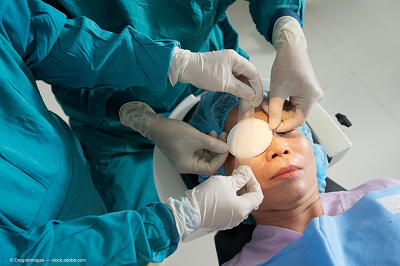
Corneal and ocular inflammation is just the tip of the proverbial iceberg, according to Victor L. Perez, MD, who noted that he views those as indicative of deeper common problems, i.e., autoimmune or systemic disorders and infections.
Know your location
Dr. Perez then advises physicians to be aware of diseases that are common in their locations, because this knowledge will help establish a diagnosis.
He provided an example based on his Boston-based training where most of the etiologies of necrotizing scleritis, including 57%, that were associated with an autoimmune disease or connective-tissue disorder.
This included systemic vasculitis disease in 48%, infection disease in 7%, and rosacea, atopy, or foreign body, in 2%.
A change of location to Bascom Palmer Eye Institute in Miami showed a different local profile, in which the most common presentation was infectious scleritis. In North Carolina, Dr. Perez said he sees several causes of necrotizing scleritis.
To treat or not to treat
An illustrative case of a scleral melt caused by a disease is that of a 60-year-old woman who presented with eye pain associated with a scleral lesion.
The patient had rheumatoid arthritis. This scenario, among others, may move physicians to the next step: to patch or not to patch. However, in Dr. Perez’s opinion, the more important consideration is to treat or not to treat.
To address that question, Dr. Perez cited two studies. Watson and Hayreh (Br J Ophthalmol 1976;60:163-91) reported that 29% of patients died within 5 years of the onset of necrotizing scleritis and 67% of those deaths resulted from extraocular vasculitis. Foster et al. (Ophthalmology 1984;10:1253-63) reported that 54% of patients who were not immunosuppressed died.
“Medical therapy, rather than surgery, is always the first line of therapy,” he said.
Dr. Perez cited a review of the expert guidelines for use of immunosuppressive drugs in patients with ocular inflammatory diseases (Am J Ophthalmol 2000;130:492-513). It looked at the patients who need immunosuppression.
“Nearly any ocular inflammatory disorder requires chronic systemic steroids,” he said. “These diseases include Behcet’s disease, necrotizing scleritis and vasculitides, birdshot retinochoroidopathy, multifocal choroiditis and panuveitis, serpiginous choroidopathy, mucous membrane pemphigoid, and sympathetic ophthalmia. Necrotizing scleritis is one of the top candidates for immunosuppression.”
Dr. Perez advised physicians to avoid jumping immediately to patching. He said it is important to first show how severe the necrotizing scleritis is and look for an infectious necrotizing scleritis.
“Do not panic,” he said. “ All scleral melts will look emergent, but surgical intervention may not be needed. Always culture the patient and biopsy if necessary.”
To patch or not to patch
When considering surgery, the inflammation or infection must first be controlled. The scleral tissue used can be patch graft or the entire sclera, the latter of which is preferable.
To prepare a patch graft, Dr. Perez likes to use a template to draw the area of scleral necrosis and create a patch graft; healthy tissue must be present in order to suture the graft.
The patch graft should be covered with conjunctiva or a mucous membrane graft. The former is the better approach, but in some cases the conjunctiva may have disintegrated.
Infectious keratitis
When treating infectious keratitis/keratitis, the potential algorithm is culturing, topical and/or systemic antibiotics and early surgery, which includes cryotherapy with conjunctival resection and antibiotic irrigation and perhaps subpalpebral lavage with an antibiotic or an antifungal with a corneal scleral patch graft.