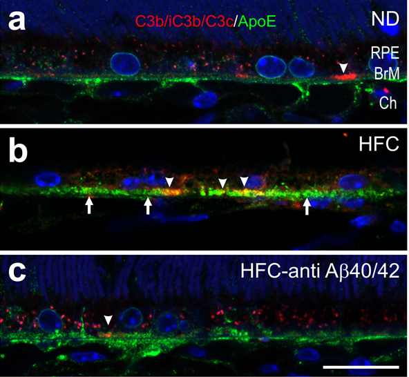
Activated complement detection in APOE4 mouse model of AMD (APOE4-HFC): Deposition of complement C3 fragments (C3b/iC3b/C3c) in APOE4 mouse eyes. (A–C) Confocal immunofluorescence images depicting immunolocalization of C3b/iC3b/C3c- (red) and ApoE- (green) positive basal deposits (arrows in B) of 75-wk-old (A) normal control APOE4-ND (ND), (B) affected APOE4-HFC (HFC), and (C) anti-Aβ40/42–treated APOE4-HFC (HFC–anti-Aβ40/42) mice. C3 fragments are found in small, sub-RPE patches (arrowheads in B). These patches are diminished in anti-Aβ40/42–treated APOE4-HFC and normal control APOE4-ND mice compared with affected APOE4-HFC animals. BrM, Bruch’s membrane; Ch, choroid; RPE, retinal pigment epithelium. (Scale bar: 20 μm.) (PMID: 21690377)
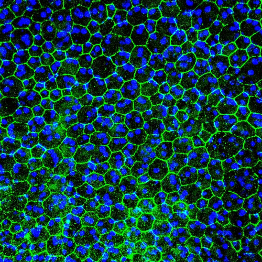
RPE flat mount of posterior mouse eye with AMD-like changes: Confocal image of RPE flat mount (apical side up) of an old mouse expressing the human complement factor H His402 risk variant (CFH H/H. Cfh-/-) and fed a high fat, cholesterol rich diet for 8 weeks. ZO-1 labeling (green) of tight junctions reveals typical "cobble stone" shape of RPE cells. Dapi staining (blue) of the nuclei reveal multinucleation (≥3 nuclei/cell) in dysmorphic RPE cells.
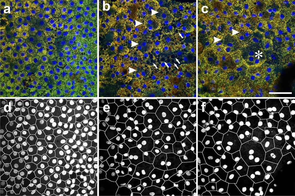
Human RPE flat mounts. (A–C) Confocal fluorescence images of RPE flat mounts from equivalent perimacular regions (RPE basal side up). (A) Seventy- six year old (Y) Caucasian males with no history of ocular disease are shown; (B and C) 85 Y Caucasian females with clinically documented dry AMD in an area with multiple binucleate RPE cells (arrowheads in B) and vacuoles (arrows in B) and an area with a trinucleate RPE cell (asterisk in C) is shown. (D–F) A–C are segmented to highlight cell borders and nuclei using DOCAP. (Scale bar: 50 μm.). (PMID: 21690377)