Capturing a Bright Future
Pioneering early detection of pediatric retinal disorders
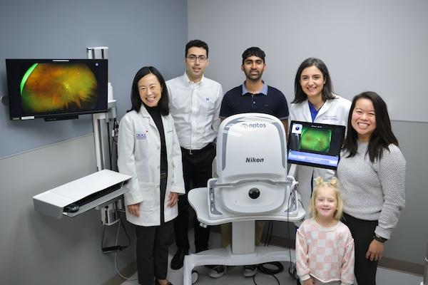
In pediatric ophthalmology, early detection of retina-related disorders is the key to unlocking a future full of possibility for young patients. Yet, all too often children do not recognize, or are unable to, articulate their vision problems and our current screening methods may not recognize retinal issues. This leads to delayed diagnoses and long-term negative impacts on their eyesight. However, a groundbreaking study — Pediatric Retina Artificial Intelligence System Evaluation (PRAISE) — is under way at Duke Eye Center and holds the potential to transform routine childhood eye exams. By adding the use of wide field fundus photography to the standard eye chart and red reflex testing, the intent is to advance the discovery of retinal detachment and other retinal problems.
Still in its initial phases, the PRAISE study shows how the laser-assisted tele-screening technology, called an optomap, is enabling ophthalmologists, optometrists, pediatricians and other professionals to capture ultra-widefield, detailed images of the fundus – the back of a child's eye – without the discomfort of traditional methods involving bright lights, dilating drops or more involved procedures that require sedation. Lejla Vajzovic, MD the pioneering force leading the study emphasizes the transformative power of this innovative approach to pediatric eye care especially as a screening tool in pediatrician and other primary are offices.
“Employing this technology as a screening tool in pediatric and primary care offices will be a game changer, giving us the ability to detect retina issues early on and prevent the need for invasive procedures,” said Vajzovic. “Through this technology, doctors are equipped to capture thorough imagery of the eye and able to make informed treatment decisions that will result in positive outcomes and better quality of life down the road for countless children.”
Important Implications
What sets the PRAISE study apart is its ambition to integrate tele-screening technology into primary care practices and school settings. The ongoing study, conducted in collaboration May Slowik, MD, a board-certified pediatrician in Durham, NC, has already imaged and estimated 150 children, with results revealing that nearly 5 percent of the patients may have undetected issues that could benefit from corrective treatment.
“It is a privilege for our primary care clinic to partner with pediatric ophthalmology to share the common goal of early childhood eye disease detection and prevention,” shared Slowik.” From toddlers to teenagers, our patients and their families have been excited to see their retinal images and to contribute to advancements in pediatric vision screening.”
In addition to the study, Vajzovic and her team are working in partnership with Duke Biomedical Engineering in the development of technology that will read the exam images and automatically deliver a pathology report. Ultimately, the hope is that by pairing the use of wide field fundus photography with artificial intelligence for automatic analysis, testing facilities of all types will be capable of providing quick assessments and facilitating timely referrals to eye specialists.
Beyond its applications in pediatric care, this tele-screening technology has already been proven successful in the care and diagnosis of patients with diabetes and among endocrinologists, showcasing its versatility and effectiveness in a variety of medical fields. The prospect of identifying retinal issues such as retinal detachment or Coats’ disease early on, opens doors for more positive outcomes and brighter futures for children.
“We are excited to pave the way for this technology to be used to revolutionize vision screenings, progress pediatric eye care and improve outcomes for children everywhere,” shared Vajzovic.