The Duke Pediatric Retina and Optic Nerve Center (DPROC) team provides unique expertise, diagnostic tools and data to ensure safe treatment of pediatric eye and brain issues.
We are dedicated to providing world-class care and quality research to pediatric patients with eye and brain conditions.
- Treating patients around the world
- Teaching residents, fellows and visiting scholars about diseases, research practices and treatments
- Researching retinal and optic nerve diseases
- Establishing a registry to catalog disease data to accelerate research and tailor clinical trials
- Providing a dedicated avenue for donor support
Spectral Domain Optical Coherence Tomography (SD-OCT) Imaging of Neural Development
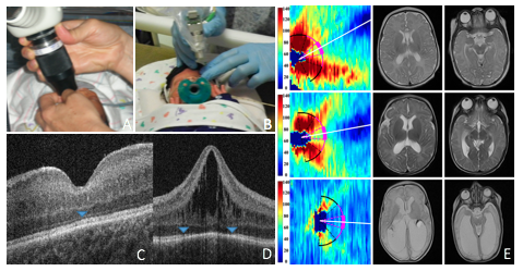
These images demonstrate the hand-held SD-OCT imaging in the NICU and Spectral domain optical coherence tomography (SD-OCT) image of preterm infant fovea.
A) Envisu 2300 Bioptigen
B) New handheld OCT device does not need a speculum and the probe does not come in contact with the infant. A pacifier and sucrose maybe used, however the infant often sleeps though the exam due to low level illumination and thus lack of associated discomfort.
C) Normal retina image at 42 weeks (blue head indicating ellipsoid zone, an indicator of photoreceptor development, at the fovea)
D) A preterm infant with macular edema (41 weeks (post-menstrual age) PMA, blue arrow heads indicating the points up to which photoreceptors are developed, a delay relative to (C)).
E) SD-OCT volumes around the optic nerve are segmented to show nerve fiber layer thickness map in three infants. Thickness at the temporal quadrant (hot pink) is 86 (top), 74 (middle) and 38µm (bottom) with corresponding near normal global brain MRI lesion burden score of 2 (top), score of 5 with mildly enlarged ventricles especially in the occipital lobe (middle) and score of 15 with markedly enlarged ventricles(bottom).