
From our Chair, Edward G. Buckley, MD

Welcome! As you flip through our 2024 VISION magazine, we’re excited to show you what’s happening at the Duke Eye Center and in the field of Ophthalmology. This year, we’ve got some truly amazing stories to share about the incredible work our team is doing. I hope you find these stories as inspiring as I do. It’s the passion and dedication of our team that makes all this possible.
-Edward Buckley, MD
Read VISION 2024
You can also read what's inside VISION 2024 by selecting the content below.


New Vice Chair Appointments for Faculty Affairs and Clinical Operations

Vice Chair for Faculty Affairs
Sharon Fekrat, MD, FACS, FASRS, professor of ophthalmology and neurology, has been appointed Vice Chair for Faculty Affairs. She is a distinguished retina specialist, and an accomplished educator and clinical researcher with broad administrative and leadership experience. Fekrat has a proven record of faculty support, mentoring, and faculty development. At Duke, she has held several leadership positions including Vice Chair of the School of Medicine Clinical Sciences Appointments, Promotions, and Tenure Committee, Co-Chair of the 5-year review of department of anesthesiology, Director of Ophthalmology Faculty Mentoring and Career Development, Director of the Vitreoretinal Surgery Fellowship, and she is founder and Director of the iMIND Research Group. She founded and spearheaded Duke Eye Center’s All About Your Eyes for patients, The Duke Manuals of Ophthalmic Surgery for eye surgeons, and Duke Journal of Case Reports in Ophthalmology for ophthalmologists. At Duke's VA affiliate (Durham VA Health Care System), she has been Chief of Ophthalmology, Interim Chief of Surgery, and is currently Associate Chief of Staff.

Vice Chair for Clinical Operations
Amy M. Fowler, MD, associate professor of ophthalmology, brings a wealth of experience to the Vice Chair for Clinical Operations. An avid Duke alumna (BS ’93, HS ’02), as a Duke resident she was an inaugural recipient of the K. Alexander Dastgheib Resident Surgery Award. In 2020, she joined the Duke Eye Center oculofacial plastic surgery division. Previously, she served 12 years as the ophthalmology residency director and served as Vice Chair for the UNC department of ophthalmology. During that time, she led the Faculty Compensation Committee and participated in the Outpatient Task Force Committee, which managed clinical operations. She has had extensive experience in a variety of clinical settings including both Duke and UNC hospitals, CMC hospitals, VA Hospitals, community and hospital based ambulatory surgery centers, and private practice clinics. Dr. Fowler has always valued the comradery and world class patient care the Duke Eye Center provides and is excited to support and help further the department’s mission of excellence in her new role.


BONUS: Research Roundup
Retinal Imaging and Alzheimer’s Disease
Advancements in Optical Coherence Tomography and Neurodegenerative Disease
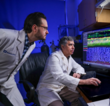
Neurodegenerative diseases are becoming ever more prevalent in our aging population. Given the debilitating and progressive nature of these diseases, early and cost-effective diagnosis of these diseases is paramount. However, conventional diagnostics such as brain magnetic resonance imaging, positron emission tomography, cerebrospinal fluid biomarkers, and genetic testing are expensive and not often readily available, which implies a more accessible alternative. Retinal imaging may be a potential alternative given that neurodegenerative changes in the brain have been associated with structural and microvascular changes in the retina, and machine learning models have been developed to distinguish retinal images using optical coherence tomography (OCT) and OCT angiography (OCTA) of patients with cognitive impairment and Alzheimer’s disease from those of healthy controls.
Optical Coherence Tomography and Eye Care
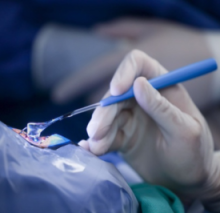
In the October 19, 2023 issue of New England Journal of Medicine article, “Optical Coherence Tomography and Eye Care,” Cynthia A. Toth, MD, professor of ophthalmology and biomedical engineering explains the invention, translation, and use of optical coherence tomography (OCT) in clinical care in ophthalmology. The article aims to communicate how and why the novel OCT technology has revolutionized eye care; andto salute James G. Fujimoto, PhD; David Huang, MD, PhD and Eric A. Swanson, PhD who were recognized with the 2023 Lasker–DeBakey Clinical Medical Research Award for their work.
Preterm Infant Retinal OCT Markers of Perinatal Health and Retinopathy of Prematurity Frontiers in Pediatrics
Optical coherence tomography (OCT) is a powerful imaging modality that has recently been adapted to the infant population and provides noninvasive, high-resolution, cross-sectional imaging of the infant eye at the bedside with low stress relative to conventional examination. In a review published in Frontiers in Pediatrics, first author, Shwetha Mangalesh, MBBS, delves into discussing the associations between preterm systemic health factors and OCT-based retinal findings and their potential contribution to the development of non-invasive biomarkers for infant health and for retinopathy of prematurity (ROP).